Galerie photos/Image-4.jpg
Précédent | Accueil | Suivant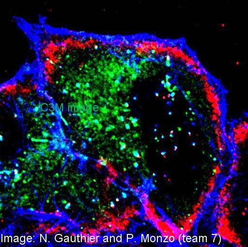
Cellules HeLa fixées (microscopie confocale): triple marquage pour CD2AP (protéine de fusion fluorescente GFP, en vert), actine-F (rhodamine phalloidine, en bleu) et VacA (anticorps, en rouge). Image: Nils Gauthier et Pascale Monzo (équipes 6 et 7) lors d'un travail collaboratif (J. Cell Biol 2007 Apr 23;177(2):343-54)
HeLa cells expressing GFP-CD2AP (green). These cells were incubated with VacA at 4°C for 1 h and were washed and incubated for 30 min at 37°C. Cells were fixed, permeabilized, and processed for the detection of actin (phalloidine, blue) and VacA (antibody, red), and observed in confocal microscopy. Image: Nils Gauthier et Pascale Monzo (teams 6 and 7) lors d'un travail collaboratif (J. Cell Biol 2007 Apr 23;177(2):343-54)